Forums » News and Announcements
Hemostatic Agents and Tissue Sealants
-
Hemostatic Agents and Tissue Sealants
OBJECTIVE. Topical tissue sealants and hemostatic agents, seen on postoperative imaging in a variety of intraabdominal and pelvic locations, have the potential to be mistaken for abdominal abnormalities, especially if the radiologist is not aware of the patient's surgical history. The normal appearance of these agents may mimic abscesses, tumors, enlarged lymph nodes, or retained foreign bodies. Therefore, it is important to be familiar with their typical imaging appearances and to review the surgical records when needed to avoid misdiagnoses. The purpose of this article is to increase the radiologist's familiarity with various types of topical tissue sealants and hemostatic agents used during surgical and percutaneous procedures in the abdomen and pelvis along with their radiologic appearances.To get more news about hemostasis, you can visit rusuntacmed.com official website.
CONCLUSION. Various types of hemostatic agents are now commonly used during surgery and percutaneous procedures in the abdomen and pelvis, and it is important to recognize the various appearances of these agents. Although there are suggestive features outlined in this article, the most important factor for the radiologist is to be aware of the patient's history and the possibility that a hemostatic agent may be present. On postoperative imaging, hemostatic agents may mimic abscesses, tumors, enlarged lymph nodes, or retained foreign bodies, and accurate diagnosis can save a patient unnecessary treatment. It is therefore crucial to incorporate knowledge of the patient's surgical history with recognition of the typical imaging appearances of hemostatic agents and other pseudolesions to avoid misdiagnoses.

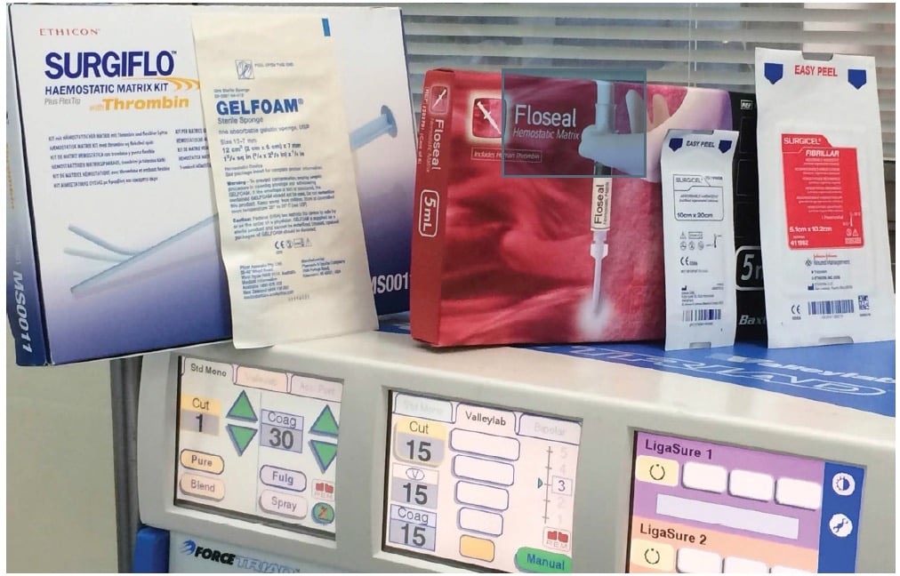
The intraoperative use of hemostatic agents and tissue sealants has increased significantly recently, largely because of the increasing trend toward minimally invasive surgeries and percutaneous interventional procedures. It is estimated that they were used in 35.2% of major surgeries in 2010 [1]. Unfortunately, with this rise in use comes new potential imaging pitfalls, because misinterpretation of the findings has been shown to be extremely common if the radiologist is not aware of the presence of these agents [2]. They can be used in a diverse array of procedures and can mimic tumor or infection in any part of the body [3, 4]. Therefore, an understanding of the agents and common imaging findings is crucial in postoperative imaging.The major types of the topical hemostatic agents include gelatin-based products, oxidized cellulose, collagen-based products, polysaccharide spheres, and fibrin sealants, the latter of which can be used in various forms as a topical hemostat, sealant, or adhesive [5–8]. There are relatively few reports of their imaging appearances after various abdominal and pelvic procedures in the literature. These agents have been most frequently described in the urologic literature, primarily after partial nephrectomies.
In this article, we discuss the imaging appearances of commonly used hemostatic agents and tissue sealants in the abdomen and pelvis with examples illustrating the most frequently encountered pitfalls. The individual agents are examined as well, although these cannot be reliably differentiated from each other on imaging. Other postoperative findings such as abdominal spacers and omental packing are briefly explored. Finally, we discuss useful ways to distinguish their normal postoperative appearance from true pathologic findings such as infection or tumor recurrence.
